The eight bones in the wrist are arranged into a “proximal row,” furthest from the fingers, and a “distal row,” closest to the fingers. When the lunate, the center bone in the proximal row, loses its blood supply, Kienböck’s disease results. 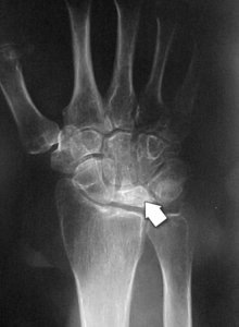
We aren’t really sure what causes this loss of blood supply, although it is associated with a shortened ulna (one of the forearm bones) and a different shape of the lunate bone itself. Trauma can affect the blood supply, but Kienböck’s really isn’t recognized as occupational.
Diagnosis of Kienböck’s is by history, physical examination, and X-rays. Most patients present with wrist pain. Sometimes, an MRI is obtained to confirm your diagnosis.
We unfortunately are not able to predict the progression of Kienböck’s. What we do know is that if and when the lunate changes so much that it fragments or collapses, the wrist joint becomes much more worn and can become more painful.
Treatments include observation, casting, and surgery. Surgery can involve changing the length of certain bones, bone grafting the lunate, or removing the lunate. In severe cases, wrist fusion might be the best option. Your results depend on severity of the disease and whether or not it progresses.